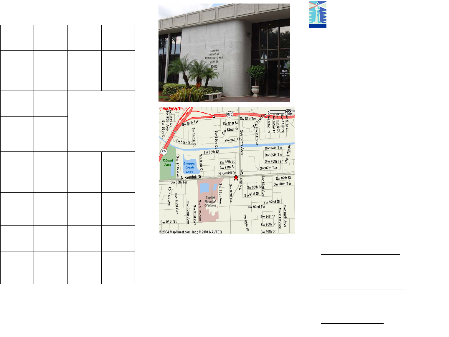
R. Wayne Whitted MD, MPH
8740 North Kendall Dr.
Suite 101
Miami, Florida 33176-2212
Phone: 305-596-3744
Fax: 305-596-3676
www.drwhitted.net
Raymond Wayne Whitted MD, MPH
...dedicated to healthy lifestyles and safe, state-of-the-art
surgery for women of all ages.
Mammography
Mammography saves lives! It is n x-ray of the
breast that is safe and is used to detect prob-
lems with a woman’s breasts. It uses a special,
low-dose x-ray machine to take pictures of
both breasts. Mammograms allow a doctor to
look closer at the breast tissue identifying
lumps and abnormalities. Mammography is
the best screening tool that doctors have for
evaluating the breast tissue.
Mammograms have both benefits and limita-
tions. For example, some cancers can’t be de-
tected by a mammogram, but may be detect-
able by breast self-exam and/or clinical breast
examination.
Checking your own breasts for lumps or other
changes is called a breast self-examination
(BSE). Studies so far have not shown that BSE
alone reduces the numbers of deaths from
breast cancer. BSE should not take the place
of clinical breast exam and a mammogram.
Are there different types of Mammo-
grams?
Screening Mammograms: are done for
women who have no symptoms of breast
cancer. The American Cancer Society rec-
ommends having a screening Mammogram
yearly after the age of 40.
Diagnostic Mammograms: are done
when a woman has symptoms of breast
cancer or a breast lump. This Mammogram
takes longer because more pictures are
taken (usually 4 for each breast).
Digital Mammogram: an electronic image
is stored on a computer. Current research
has not shown that digital images are bet-
ter at finding cancer than x-ray images.
American College of Radiology BI-RADS Categories
ACR
Category
(Birads)
Assessment Probability
of
cancer
% of MMG
0 Needs addi-
tional imag-
ing or com-
parison films
NA < 10%
1 Negative;
routine fol-
low-up
NA
2 Benign find-
ings/negativ
e; routine
follow-up
3 Probably
benign:
short inter-
val f/u sug-
gested
<2% 4%
4 Suspicious
abnormality:
bx suggested
3-49% 2/1000-
8/1000
5 Highly sug-
gestive of
malignancy
>95%
6 Biopsy
proven ma-
lignancy
NA NA
>90%
What if a lump is found?
Your doctor may order other tests, such as an
ultrasound to determine if the lump is cystic or
solid. Occasionally a biopsy, a test where a
small amount of tissue is taken from the lump
and area around the lump, may be ordered. The
tissue is sent to a lab to look for cancer or
changes that may mean cancer is likely to de-
velop.
Breast lumps or growths can be benign (not
cancer) or malignant (cancer). Finding breast
cancer early means that a woman has a better
chance of surviving the disease. There are also
more choices for treatment when breast cancer
is found early.
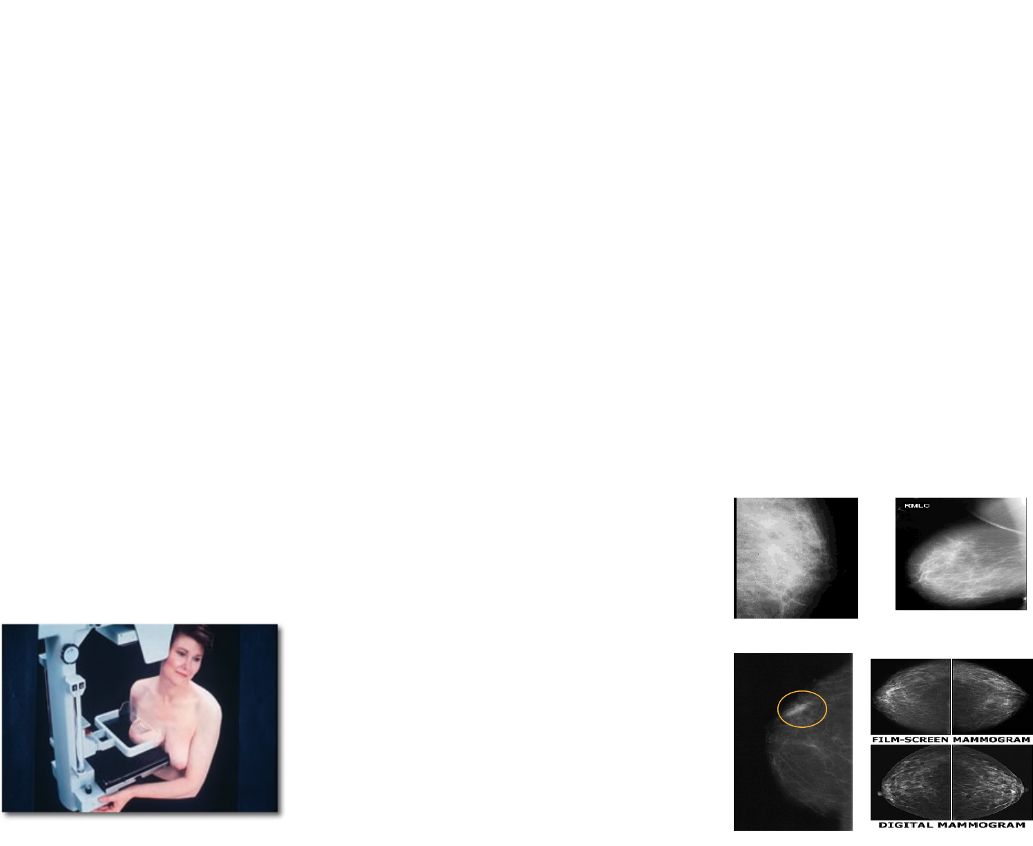
How is a Mammogram done?
You stand in front of a special x-ray machine.
The x-ray technologist places your breasts (one
at a time between two plastic plates. The plates
press your breast to make it flat so that the x-
ray can penetrate the breast tissue and make
the test more accurate. This may cause you
some discomfort such as a squeezing or pinch-
ing. It is better to have your mammogram just
after your menstrual period.
Most often, two pictures are taken of each
breast-one from the side and one from above. A
screening mammogram takes about 15 minutes
to accomplish.
What if I have breast implants?
If you have breast implants, be sure to tell your
mammography facility when you make an ap-
pointment. You will need an x-ray tech who is
trained in dealing with implants. This is impor-
tant because breast implants can hide some
breast tissue, which could make it difficult for
the radiologist to see breast cancer when look-
ing. For this reason, to take a mammogram of
breast with an implant, the technician might
gently lift the breast tissue slightly away from
the implant.
How often should I get a mammogram?
The American Cancer Society guidelines
states a woman should get yearly mammo-
grams beginning at age 40.
Women who have had breast cancer or
other breast problems or who have a family
history of breast cancer might need to start
getting mammograms before age 40 or they
might need to get them more often than
yearly. Talk with your doctor about when to
start and how often you should have a
mammogram.
How do I get ready for my mammogram?
Schedule your mammogram just after your
period because your breasts are less ten-
der.
Take an anti-inflammatory medicine like ad-
vil before you have your procedure.
Acknowledge breast implants when you
make your appointment if you have them.
Wear a shirt with shorts, pants, or skirt so
that you only have to remove your top.
Don’t wear any deodorant, perfume, lotion,
or powder under your arms or on your
breasts on the day of your mammogram.
These things may make shadows on you x-
ray picture.
...
dedicated to healthy lifestyles and safe, state-of-the-art, innovative surgery for women of all ages
Are there any problems with mammo-
grams?
The limits of mammography include:
They are only part of a complete exam.
Your doctor and your radiologist should do a
clinical breast exam.
False negatives: this means everything
may look normal on the mammogram when
in fact something abnormal is present.
Younger women and women on meno-
pause hormone therapy are more likely to
have this because of denser breast tissue.
The average reported rate for mammogra-
phy is 10% (1 out of 10)
False positives: This is when the mammo-
gram results look like an abnormality when
in fact there is no abnormality. False posi-
tives are also more common in younger
women and those who take menopausal
hormone therapy. These may lead to many
unnecessary biopsies.
Examples of Mammogram Pictures
Dense Breasts Non-Dense Breast
Breast Cancer Mammogram types